Connect with us

CivicSciTimes - Stories in Science

My Passion for Microbes

Dr. Chika Ejikeugwu is a Lecturer at Ebonyi State University, Abakaliki in Nigeria where he teaches microbiology to undergraduate students. He is also an ‘associate’ Development Knowledge Facilitator (DKF) for the National Youth Service Corp (NYSC) headquarters, Abuja, Nigeria. He holds a doctorate degree in Pharmaceutical Microbiology & Biotechnology from Nnamdi Azikiwe University, Awka, Nigeria.

CSM Lab

Published

on

Chika Ejikeugwu

[su_boxbox title=”About”]Dr. Chika Ejikeugwu is a Lecturer at Ebonyi State University, Abakaliki in Nigeria where he teaches microbiology to undergraduate students. He is also an ‘Associate’ Development Knowledge Facilitator (DKF) for the National Youth Service Corp (NYSC) headquarters, Abuja, Nigeria. He holds a doctorate degree in Pharmaceutical Microbiology & Biotechnology from Nnamdi Azikiwe University, Awka, Nigeria. The edited story below was originally published by Science Communication Hub Nigeria. [/su_boxbox]

[dropcap]A[/dropcap]s far back as I can remember, I have always wanted to be a scientist. Of course, I also thought about becoming a medical doctor as well from time to time. However, after gaining admission to study microbiology at university, I dropped all thoughts about studying medicine because microbiology opened my eyes to a brand-new world of microbes. These little things provided me with a new way I could impact society.

In 2015, I created and founded an online platform called MicroDok for the teaching and study of microbiology. The site is now the number one and largest microbiology website in Nigeria and Africa. It has also attracted many followers from around the world. After completing my doctoral thesis in July 2017, I received the 2018 Matsumae International Foundation (MIF) postdoctoral fellowship award to study at Kyoto University in Japan. There, I studied HIV-1 for 6 months. I was 32 years at that time, and also the youngest fellow to receive that award from Nigeria and around the world.

Broadly, my research interests are currently clustered around understanding the molecular mechanisms behind antibiotic resistance, and functioning HIV-1. I thoroughly enjoy research especially given that the topics I am studying are global problems to which Nigeria is not immune. For example, with respect to antibiotic resistance, I am energized because it cuts across all fields of medicine since we cannot fight infectious diseases without antibiotics. However, the efficacy of these antibiotics is being threatened by some resistant microbes. Microbiology is an exciting field that literally impacts may facets of life. I am captivated with how microbes – as invisible as they are to the naked eye – can positively transform society.

Explore Next:  Science Inspiration and Breaking the Mold - Mixing Fashion and Science

The challenges facing scientists in Nigeria are numerous. This includes lack of funding, lack of collaboration amongst Nigerian scientists, epileptic power supply, and dilapidating infrastructure. These are challenges that many scientists in Nigeria face. Nonetheless, we are passionate about the work which keeps us moving forward despite the challenges.

I remain excited about my research. I am hopeful to take up the Alexander von Humboldt Postdoctoral Fellowship in Berlin, Germany to do another round of research on antibiotic resistance for two years. I plan to also sustain my collaboration with Kyoto University on my HIV-1 research. I continue to explore ways my current university in Nigeria could establish a student and staff exchange program in an effort to help strengthen the academic and research profile of my home University.

My advice for the aspiring scientist is to remain resilient. The journey in science is tough. It is especially tough doing science in Nigeria and Africa at large. But I strongly believe that with preparation, determination, focus, a little help from mentors, and a bit of luck, every mountain can be overcome.

Cover image from Science Communication Hub Nigeria

Metrics

Sessions

[analytify-stats metrics=”ga:sessions” analytics_for=”current” custom_page_id = “”permission_view=””]

Total number of Sessions. A session is the period time a user is actively engaged with the page.

Visitors

[analytify-stats metrics=”ga:visitors” analytics_for=”current” custom_page_id = “”permission_view=””]

Users that have had at least one session within the selected date range. Includes both new and returning users.

Page views

[analytify-stats metrics=”ga:pageviews” analytics_for=”current” custom_page_id = “”permission_view=””]

Pageviews is the total number of time the article was viewed. Repeated views are counted.

CivicSciTimes - Stories in Science

Unexpected Stories and Spindle Mistakes: Discovering that Wild-type Cells are Full of Surprises

CSM Lab

Published

on

By

Natalie Nannas

Natalie Nannas is an Associate Professor of Biology at Hamilton College in Clinton, NY. She teaches courses in genetics, molecular biology, and bioethics. Dr. Nannas graduated from Grinnell College with bachelor’s degrees in biological chemistry and French. She received her Master’s and PhD from Harvard University in molecular biology and genetics. Dr. Nannas conducted her postdoctoral research at the University of Georgia where she won a National Science Foundation Plant Genome Postdoctoral Fellowship. At Hamilton College, Dr. Nannas enjoys teaching and sharing her passion for microscopy with her undergraduate research students. When not glued to a microscope, she loves spending time with her husband and two daughters. The narrative below by Natalie Nannas captures the human stories behind the science from a 2022 paper titled “Frequent spindle errors require structural rearrangement to complete meiosis in Zea mays” which was published by her group in 2022 in the International Journal of Molecular Sciences.

Science never works out the way we plan. As scientists, we ask questions, hypothesize and outline our goals … then reality of science occurs. The reality of science is often full of failed controls, endless troubleshooting, and sometimes strange findings that lead us in new and unpredictable directions. Our publications give the impression that we planned these scientific journeys from the beginning and do not tell the human side of the process with all of its twists and turns, dead-ends and U-turns. I want to tell you the real story behind my first publication as a faculty member with my own lab. It did not go as planned due to the COVID-19 pandemic. My lab was shut down in the middle of our investigation, and my students and I were unable to generate new data. In the beginning, it seemed like we were stranded with only control data and no story to tell, but the time away from the lab allowed us to spend more time looking carefully at wild-type cells. What seemed like a dead-end suddenly became its own story when we found something unexpected hiding within microscopy movies. Our wild-type cells were making mistakes, attempting fixes and changing directions, just like we do as scientists.

My scientific journey began with flickering green lights and a microscope (you can read more about it here). As an undergraduate, I was mesmerized by the beauty of watching living cells shuffle fluorescently labeled proteins throughout their cytoplasm. I followed this passion for microscopy into my doctoral dissertation research at Harvard University where I investigated how yeast cells build the machinery needed to pull their chromosomes apart. This machinery is a dynamic collection of long protein tubes called microtubules and other organizing proteins that help move and shuffle microtubules. I loved watching the delicate dance of chromosomes interacting with microtubules of the spindle, and I wanted to continue studying this process in my postdoctoral studies.

During postdoctoral studies at the University of Georgia, I won a fellowship from the National Science Foundation to develop a new technique in microscopy. No one had ever watched plants building their spindles in meiosis, the specialized cell division that produces egg and sperm. Other scientists had performed beautiful microscopy studies observing how mitotic spindles function inside of plant cells, but due to the technical challenges, no one had ever observed live plant cells building spindles in meiosis. I was thrilled to take on this challenge by using version of maize that had fluorescently labeled tubulin, the protein that makes up microtubules of the spindle. With this line of maize, spindles would glow fluorescent green, allowing me to image if only I could extract the meiotic cells.

Dr. Natalie Nannas

We were so busy collecting data and prepping for our mutant studies that we never really took time to analyze the wild-type cells.

After almost a year spent dissecting maize plants, I finally managed to develop a method to isolate these tiny cells and keep them alive in a growth media long enough to image them. This new method of live imaging was going to serve as the foundation of my new lab at Hamilton College, a primarily undergraduate institution. With my students, I planned to investigate the pathways governed spindle assembly. Most animal mitotic cells have a structure called a centrosome that dictates how spindles are formed; however, female animal meiotic cells lack these structures and must use other pathways to direct spindle assembly. Plants also lack centrosomes, and I wanted to inhibit these known animal pathways in our plant live imaging system.

Explore Next:  Unexpected lessons from conducting research with older adults

As I set up my lab, my students and I collected live movies of wild-type maize cells building their spindles. I told my students and myself that these movies were not the main event, they were just the control cells so we would have a baseline comparison for our experimental conditions. We were so busy collecting data and prepping for our mutant studies that we never really took the time to analyze the wild-type cells. At the surface level, they built spindles and segregated chromosomes in a generally expected amount of time, so we focused on preparing for our upcoming experiments…. then March 2020 occurred.

The pandemic forced us to slow down and look more carefully at our wild-type data, and I am grateful for the detour.

My students headed home for spring break with a warning that there may be a delay in coming back to campus due to the spread of COVID-19. None of us were prepared for the shutdown that followed. Like many colleges and universities, our campus was closed for the remainder of the spring 2020 semester and the summer of 2020. My students and I began meeting on Zoom, trying to make a new plan for our research. The only data we had to work with were the microscopy of wild-type maize cells, so we decided to spend time digging more deeply into these movies. Originally, we had only measured the total time it took to build a spindle as it would be a baseline for comparison to our mutants. We had not looked carefully at any of the intermediate time points in the assembly process. When my students looked more closely at our movies, they discovered that wild-type cells built an incorrectly shaped spindle over 60% of the time!

We found that maize meiotic cells often built spindles with three poles instead of two, and they had to actively rearrange their spindle structure to correct this mistake. We also found that in these cells, there was a delay in meiosis as cells refused to progress until this correction had been made. This is an exciting discovery as it showed that plants are error-prone in their spindle assembly, much like human female meiotic cells. Our findings also suggested that meiotic cells were monitoring their spindle shape when determining if they should move forward in meiosis. Previous work has shown that cells monitor the attachment of chromosomes to the spindle to make this decision, but our work adds a new dimension, showing that they also monitor spindle shape. As we continued to analyze our videos, we also learned that cells corrected their spindle morphology in a predictable way. They always collapsed the two poles that were closest together, creating a single pole and resulting in a correct bipolar spindle.

The image shows the first page of the paper which can be accessed here.

My students and I had begun our scientific journey planning to breeze over wild-type cells, moving on to what we envisioned would be a more exciting story of spindle mutants. The pandemic forced us to slow down and look more carefully at our wild-type data, and I am grateful for the detour. I rediscovered my love of closely watching flickering green fluorescent lights, the dance of microtubules sliding into place or making missteps and shuffling into new arrangements. Watching life attempt a complicated process, make mistakes, and try again, is a lesson that never grows old. It reminds me that our scientific journeys are just the same, they start in one direction but are fluid and constantly changing, and hopefully, they end with a functional spindle!

Read the Published Paper

Weiss, J.D., McVey, S.L., Stinebaugh, S.E., Sullivan, C.F., Dawe, R.K., and N.J. Nannas. 2022. Frequent spindle errors require structural rearrangement to complete meiosis in Zea maysInternational Journal of Molecular Sciences, 23 (8):4293–4312.

ABOUT: Stories in Science is a special series on the Civic Science Times. The main aim is to document the first-hand accounts of the human stories behind the science being published by scientists around the world. Such stories are an important element behind the civic nature of science.
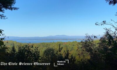
SUBMISSION: Click here to access the story guidelines and submission portal. Please note that not all stories are accepted for publication. After submission, we will let you know whether we have selected the story for the review process.

Continue Reading

Upcoming Events

Trending