Connect with us

CivicSciTimes - Stories in Science

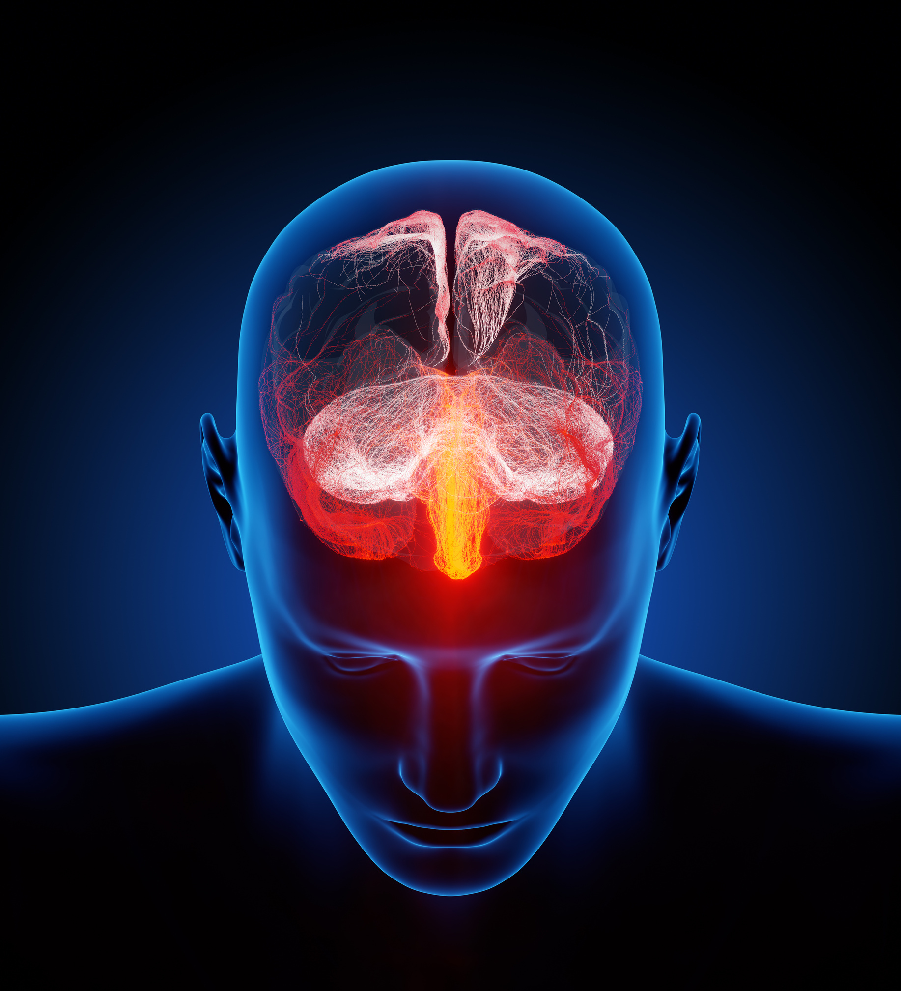
From engrams to psychiatric disorders and back

CSM Lab

Published

on

– Steve Ramirez –  

– Principal Investigator | Center for Brain Science| Harvard University – 

[dropcap]I[/dropcap]‘m often asked how I got into neuroscience, so here’s the story. In college, I was one of those students who loved every subject, from Shakespeare to biochemistry to physics — which is a roundabout way of saying I had no idea what I wanted to do with my life. I was volunteering in a lab just to see what research really entailed, and one day a centrifuge broke. I went to the lab across the hall to ask if we could use theirs and, serendipitously, a girl who I had a crush on sophomore year was standing there using it. Naturally, I started sweating and thinking of one billion ways to open up a conversation without instantly passing out, and centrifuge dynamics seemed like the most romantic topic at the moment. We started chatting about our lack of majors, classes taken, etc, and given my broad interests, she pointed me in the direction of a guy who she thought might help — he would soon become the director of Boston University’s undergraduate neuroscience program (which didn’t exist at the time).

I learned that research was incredibly hard, incredibly rewarding, and incredibly social.

steve ramirez

After one conversation with him, it clicked. “Why not study the organ that’s produced everything that’s ever been produced?” he asked. From Hamlet to vaccinations to satellites flying past Pluto, all of these achievements were the product of the most multi-disciplinary organ known: the brain. It was the common denominator across all my interests. Within a few weeks, I began working in Howard Eichenbaum’s lab with Chris MacDonald studying time cells and memory, and research quickly had me hooked. The lab’s high-octane enthusiasm and collaborative approach to science was contagious and inspiring. I learned that research was incredibly hard, incredibly rewarding, and incredibly social. I was all in with neuroscience.

In my opinion, it was a privilege to discover for a living; and, each day felt more like a roundhouse kick to the adrenal glands

However, I was never a 4.0 GPA person and according to my GRE scores, I can barely speak english or do long division, but that’s why research made sense to me: these numbers don’t matter in a world of asking questions, testing hypotheses, and observing brains in action. All that matters here is hard work and good luck. Given my test scores, it still feels like a miracle that MIT decided to accept me into their program. From day one onward, graduate school simultaneously was as humbling as it was invigorating. In my opinion, it was a privilege to discover for a living; and, each day felt more like a roundhouse kick to the adrenal glands (in the best way) and less like a stiff formal job.

Explore Next:  How to Create a Supportive Space for Scientists to Share Stories of Struggles

I went on to earn my Ph.D. in Susumu Tonegawa’s lab at MIT by working alongside Xu Liu and studying the cellular basis of memories. I worked with a team that I fully admire, aspire to represent, and am endlessly thankful for. Our team was full of camaraderie, mutual respect, wide-eyed ambition, and manhattans — the stuff of science indeed. And finally, today, with Joshua Sanes’ endless mentorship, I am a Junior Fellow and Principal Investigator at Harvard’s Center for Brain Science studying the cellular and circuit-wide basis of memory, as well as how to leverage artificially modulated memories to alleviate maladaptive states. For all this, I have a centrifuge to thank.


The Ramirez Group

Steve Ramirez’s Ted Talk 

Can This Guy Zap Away Your Bad Memories?


Featured Image (original) is by Ars Electronica and is titled “Human brain illustrated with millions of small nerves” | From Flickr | Some rights reserved


CivicSciTimes - Stories in Science

Unexpected Stories and Spindle Mistakes: Discovering that Wild-type Cells are Full of Surprises

CSM Lab

Published

on

By

Natalie Nannas

Natalie Nannas is an Associate Professor of Biology at Hamilton College in Clinton, NY. She teaches courses in genetics, molecular biology, and bioethics. Dr. Nannas graduated from Grinnell College with bachelor’s degrees in biological chemistry and French. She received her Master’s and PhD from Harvard University in molecular biology and genetics. Dr. Nannas conducted her postdoctoral research at the University of Georgia where she won a National Science Foundation Plant Genome Postdoctoral Fellowship. At Hamilton College, Dr. Nannas enjoys teaching and sharing her passion for microscopy with her undergraduate research students. When not glued to a microscope, she loves spending time with her husband and two daughters. The narrative below by Natalie Nannas captures the human stories behind the science from a 2022 paper titled “Frequent spindle errors require structural rearrangement to complete meiosis in Zea mays” which was published by her group in 2022 in the International Journal of Molecular Sciences.

Science never works out the way we plan. As scientists, we ask questions, hypothesize and outline our goals … then reality of science occurs. The reality of science is often full of failed controls, endless troubleshooting, and sometimes strange findings that lead us in new and unpredictable directions. Our publications give the impression that we planned these scientific journeys from the beginning and do not tell the human side of the process with all of its twists and turns, dead-ends and U-turns. I want to tell you the real story behind my first publication as a faculty member with my own lab. It did not go as planned due to the COVID-19 pandemic. My lab was shut down in the middle of our investigation, and my students and I were unable to generate new data. In the beginning, it seemed like we were stranded with only control data and no story to tell, but the time away from the lab allowed us to spend more time looking carefully at wild-type cells. What seemed like a dead-end suddenly became its own story when we found something unexpected hiding within microscopy movies. Our wild-type cells were making mistakes, attempting fixes and changing directions, just like we do as scientists.

My scientific journey began with flickering green lights and a microscope (you can read more about it here). As an undergraduate, I was mesmerized by the beauty of watching living cells shuffle fluorescently labeled proteins throughout their cytoplasm. I followed this passion for microscopy into my doctoral dissertation research at Harvard University where I investigated how yeast cells build the machinery needed to pull their chromosomes apart. This machinery is a dynamic collection of long protein tubes called microtubules and other organizing proteins that help move and shuffle microtubules. I loved watching the delicate dance of chromosomes interacting with microtubules of the spindle, and I wanted to continue studying this process in my postdoctoral studies.

During postdoctoral studies at the University of Georgia, I won a fellowship from the National Science Foundation to develop a new technique in microscopy. No one had ever watched plants building their spindles in meiosis, the specialized cell division that produces egg and sperm. Other scientists had performed beautiful microscopy studies observing how mitotic spindles function inside of plant cells, but due to the technical challenges, no one had ever observed live plant cells building spindles in meiosis. I was thrilled to take on this challenge by using version of maize that had fluorescently labeled tubulin, the protein that makes up microtubules of the spindle. With this line of maize, spindles would glow fluorescent green, allowing me to image if only I could extract the meiotic cells.

Dr. Natalie Nannas

We were so busy collecting data and prepping for our mutant studies that we never really took time to analyze the wild-type cells.

After almost a year spent dissecting maize plants, I finally managed to develop a method to isolate these tiny cells and keep them alive in a growth media long enough to image them. This new method of live imaging was going to serve as the foundation of my new lab at Hamilton College, a primarily undergraduate institution. With my students, I planned to investigate the pathways governed spindle assembly. Most animal mitotic cells have a structure called a centrosome that dictates how spindles are formed; however, female animal meiotic cells lack these structures and must use other pathways to direct spindle assembly. Plants also lack centrosomes, and I wanted to inhibit these known animal pathways in our plant live imaging system.

Explore Next:  Flickering Lights in the Darkness: How Microscopy Shaped My Scientific Path

As I set up my lab, my students and I collected live movies of wild-type maize cells building their spindles. I told my students and myself that these movies were not the main event, they were just the control cells so we would have a baseline comparison for our experimental conditions. We were so busy collecting data and prepping for our mutant studies that we never really took the time to analyze the wild-type cells. At the surface level, they built spindles and segregated chromosomes in a generally expected amount of time, so we focused on preparing for our upcoming experiments…. then March 2020 occurred.

The pandemic forced us to slow down and look more carefully at our wild-type data, and I am grateful for the detour.

My students headed home for spring break with a warning that there may be a delay in coming back to campus due to the spread of COVID-19. None of us were prepared for the shutdown that followed. Like many colleges and universities, our campus was closed for the remainder of the spring 2020 semester and the summer of 2020. My students and I began meeting on Zoom, trying to make a new plan for our research. The only data we had to work with were the microscopy of wild-type maize cells, so we decided to spend time digging more deeply into these movies. Originally, we had only measured the total time it took to build a spindle as it would be a baseline for comparison to our mutants. We had not looked carefully at any of the intermediate time points in the assembly process. When my students looked more closely at our movies, they discovered that wild-type cells built an incorrectly shaped spindle over 60% of the time!

We found that maize meiotic cells often built spindles with three poles instead of two, and they had to actively rearrange their spindle structure to correct this mistake. We also found that in these cells, there was a delay in meiosis as cells refused to progress until this correction had been made. This is an exciting discovery as it showed that plants are error-prone in their spindle assembly, much like human female meiotic cells. Our findings also suggested that meiotic cells were monitoring their spindle shape when determining if they should move forward in meiosis. Previous work has shown that cells monitor the attachment of chromosomes to the spindle to make this decision, but our work adds a new dimension, showing that they also monitor spindle shape. As we continued to analyze our videos, we also learned that cells corrected their spindle morphology in a predictable way. They always collapsed the two poles that were closest together, creating a single pole and resulting in a correct bipolar spindle.

The image shows the first page of the paper which can be accessed here.

My students and I had begun our scientific journey planning to breeze over wild-type cells, moving on to what we envisioned would be a more exciting story of spindle mutants. The pandemic forced us to slow down and look more carefully at our wild-type data, and I am grateful for the detour. I rediscovered my love of closely watching flickering green fluorescent lights, the dance of microtubules sliding into place or making missteps and shuffling into new arrangements. Watching life attempt a complicated process, make mistakes, and try again, is a lesson that never grows old. It reminds me that our scientific journeys are just the same, they start in one direction but are fluid and constantly changing, and hopefully, they end with a functional spindle!

Read the Published Paper

Weiss, J.D., McVey, S.L., Stinebaugh, S.E., Sullivan, C.F., Dawe, R.K., and N.J. Nannas. 2022. Frequent spindle errors require structural rearrangement to complete meiosis in Zea maysInternational Journal of Molecular Sciences, 23 (8):4293–4312.

ABOUT: Stories in Science is a special series on the Civic Science Times. The main aim is to document the first-hand accounts of the human stories behind the science being published by scientists around the world. Such stories are an important element behind the civic nature of science.

SUBMISSION: Click here to access the story guidelines and submission portal. Please note that not all stories are accepted for publication. After submission, we will let you know whether we have selected the story for the review process.

Continue Reading

Upcoming Events

Trending